Are you struggling with persistent foot pain? You’re not alone; it’s a common issue affecting millions. Understanding the underlying causes is crucial for effective treatment, and diagnostic imaging plays a key role.
This article explores the causes of joint foot pain and how imaging techniques like X-rays, MRIs, and CT scans help accurately diagnose it. Conditions such as plantar fasciitis, arthritis, and stress fractures can all benefit from these insights, allowing healthcare providers to create targeted treatment plans.
Don’t let foot pain hold you back. Learn how diagnostic imaging can lead to the answers and relief you deserve.
Common Causes Of Foot Pain
Foot pain can arise from several conditions, and consulting a foot specialist is essential for proper diagnosis and treatment. One common cause is plantar fasciitis, which leads to sharp heel pain, particularly in the morning, due to plantar fascia inflammation. Risk factors for this condition include obesity and wearing unsupportive footwear. Arthritis is another contributor, affecting the foot joints and causing pain and stiffness. Osteoarthritis results from wear and tear over time, while rheumatoid arthritis is driven by inflammation. Stress fractures, tiny cracks in the bone caused by repetitive stress, are also common, particularly among athletes, and lead to localized pain and swelling. Identifying these conditions is crucial for determining the most effective treatment plan.
Understanding The Importance Of Diagnostic Imaging
Diagnostic imaging is vital for diagnosing foot pain, providing insights beyond physical examinations. Techniques like X-rays, MRIs, CT scans, and ultrasounds help identify fractures, tears, and abnormalities in the foot.
Imaging can confirm or rule out conditions quickly, guiding treatment decisions. For instance, an MRI may indicate a torn ligament, suggesting physical therapy, while a severe tear might require surgery. This information significantly shapes management plans, ensuring patients receive tailored care for their conditions.
X-Ray Imaging For Foot Pain Diagnosis
X-ray imaging is a standard first-line tool for evaluating foot pain. It effectively identifies fractures, dislocations, and bone abnormalities. It uses electromagnetic radiation to produce clear images of the foot’s internal structures, allowing healthcare providers to rule out bone-related issues quickly.
This non-invasive and cost-effective method is particularly useful in trauma cases, providing immediate insights for prompt treatment. However, X-rays have limitations; they may not detect soft tissue injuries like ligament tears or tendon inflammation. If X-ray results are inconclusive, further imaging techniques may be needed for a comprehensive diagnosis.
MRI Imaging For Foot Pain Diagnosis

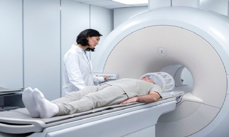
Magnetic Resonance Imaging (MRI) is crucial for diagnosing soft tissue injuries in the foot. It provides detailed images of muscles, tendons, ligaments, and cartilage. Unlike X-rays, MRIs use a magnetic field and radio waves, making them ideal for identifying conditions such as tendon ruptures and ligament tears.
MRIs can also reveal changes in bone marrow, indicating issues like stress fractures or infections. This ability to visualize both soft and hard tissues enhances diagnostic accuracy, making MRI an essential tool in managing foot pain.
CT Scan Imaging For Foot Pain Diagnosis
Computed Tomography (CT) scans are valuable for diagnosing foot pain, especially when detailed images of bone structures are required. By combining multiple X-ray images from different angles, CT scans create cross-sectional views that offer a more precise picture than standard X-rays. CT scans excel at identifying complex fractures and bone deformities that may not be visible on traditional X-rays. They are beneficial for patients with significant trauma or chronic pain of unclear origin, aiding in treatment planning and pre-surgical visualization. According to Tellica Imaging, CT scans are typically reserved for cases where additional detail is essential due to higher radiation exposure. Physicians carefully consider the risks and benefits before recommending them.
Ultrasound Imaging For Foot Pain Diagnosis
Ultrasound imaging is an increasingly popular diagnostic tool for foot pain. It utilizes high-frequency sound waves to create images of soft tissues. It’s particularly effective for evaluating conditions involving tendons, ligaments, and muscles. Being noninvasive and radiation-free, ultrasound is a safe option for many patients.
This technique is invaluable for diagnosing tendonitis, bursitis, and nerve entrapments. It allows healthcare providers to visualize foot movement and assess structural interactions during activity. Additionally, ultrasound can guide interventions such as pain relief injections or fluid aspirations, offering real-time imaging that enhances diagnosis and treatment.
The Benefits Of Using Imaging For Foot Pain Diagnosis
Integrating imaging techniques in diagnosing foot pain offers clients and healthcare providers. Critical benefitsmaging provides precise, objective data that helps clinicians make informed decisions, reduce misdiagnoses, and ensure tailored care.
Early intervention is also facilitated, allowing for prompt treatment of issues like stress fractures or soft tissue injuries, which helps prevent complications and preserves quality of life.
Finally, imaging enhances communication by allowing patients to visualize their conditions. This leads to better understanding and engagement in their treatment plans, ultimately improving outcomes.
When To Seek Medical Help For Foot Pain
It’s important to know when to seek medical help for foot pain. Consult a healthcare provider if pain persists despite rest, ice, or over-the-counter medications or if you notice swelling, bruising, or deformity.
Individuals with diabetes or chronic conditions should be extra vigilant, as they are at higher risk for complications. Symptoms like fever, redness, or warmth may indicate an infection and require immediate attention.
If foot pain affects your daily activities, seek help to receive a thorough evaluation and appropriate treatment plan.
Conclusion And Final Thoughts
In conclusion, foot pain can stem from conditions like plantar fasciitis, arthritis, and stress fractures, each requiring tailored diagnosis and treatment. Understanding these causes is essential for seeking appropriate care. Diagnostic imaging, including X-rays, MRIs, CT scans, and ultrasounds, is crucial for accurate diagnoses and effective treatment plans.
Early intervention is vital to prevent complications and improve outcomes, so it’s important to recognize when to seek medical help. Don’t let foot pain hinder your active life; by understanding your condition and utilizing diagnostic imaging, you can collaborate with your healthcare provider to achieve the relief you need. Remember, caring for your feet is critical to your overall well-being.


